
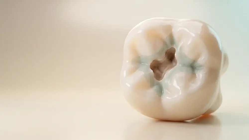
Dental decay can begin before there is a toothache and minerals may already be disappearing from the tooth’s surface before a hole appears. Also called dental caries, dental decay is a progressive disease affecting the mineralised tissues of the teeth. A cavity may appear after tooth structure breaks down.
The earliest stage of dental decay may sometimes be stopped or reversed. Once a cavity has formed, however, the missing tooth structure cannot naturally grow back and requires professional treatment.
What Is Dental Decay?
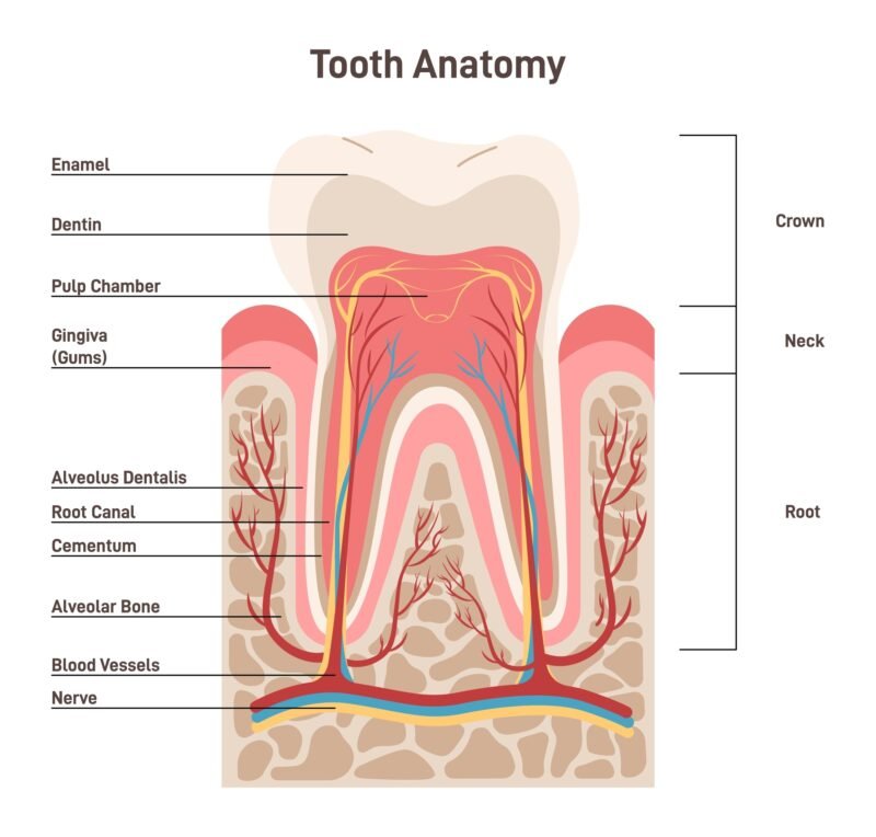
Dental decay is a biofilm-mediated, sugar-driven disease. Bacteria living in dental plaque use sugars and other carbohydrates to produce acids. These acids remove minerals, primarily calcium and phosphate, from tooth enamel. This is called demineralisation.
The teeth experience a continuous balance between mineral loss and repair. Saliva, for example, helps neutralise the acids and return minerals to the enamel through remineralisation. A cavity does not usually develop because of one sweet snack. The problem occurs when repeated acid attacks cause mineral loss to happen faster than the mouth can replace it.
The General Dental Decay Process
If plaque is not treated, it may eventually harden into dental calculus, commonly called tartar. Tartar does not directly create a cavity, but it can make plaque removal more difficult. It requires professional cleaning, as brushing and flossing cannot remove it.
- Dental plaque accumulates on a tooth.
- Plaque bacteria process sugars and fermentable carbohydrates.
- The bacteria produce acids.
- These acids remove minerals from the enamel.
- Repeated attacks weaken the tooth surface.
- A carious lesion —an area affected by dental decay— develops.
- Continued damage may create a cavity and reach deeper tissues.
How Does a Tiny Area of Dental Decay Become a Serious Problem?
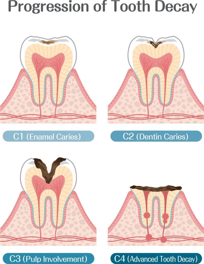
Dental caries is progressive, but not every lesion reaches an advanced stage. Early identification and appropriate care may stop some lesions from progressing. Early dental decay is often painless because enamel does not contain nerves. Pain is therefore not a reliable indication of how much damage is present.
Stage | What is happening? | What might you notice? | Can it be reversed? | Possible management |
Acid attack | Acids temporarily remove minerals from enamel | Usually nothing | Yes | Daily preventive care |
Initial demineralisation | Mineral loss exceeds natural repair, but the surface remains intact | A chalky white spot | Sometimes | Fluoride, plaque control and dietary changes |
Enamel caries | Damage progresses through the outer enamel | White, brown or dark changes; often no pain | Not after surface breakdown | Preventive care or restoration |
Dentine caries | Decay reaches the softer dentine | Sensitivity, food trapping or a visible hole | No | Usually a filling or another restoration |
Pulp involvement | Inflammation or infection reaches the nerves and blood vessels | Persistent pain, lingering sensitivity or pain when biting | No | Root canal treatment or extraction |
Dental abscess | Infection spreads around the root and pus may collect | Throbbing pain, swelling, fever or a bad taste | No | Urgent professional treatment |
Where Can Cavities Develop?
Cavities can begin on almost any exposed tooth surface. Once decay reaches softer dentine or an exposed root surface, it may progress more readily than decay limited to enamel.
Type of cavity | Where it develops | Why it may be difficult to detect or prevent |
Pit and fissure cavity | Grooves on the chewing surfaces of back teeth | Narrow grooves can trap plaque and food |
Smooth-surface cavity | Flat sides of teeth, including between neighbouring teeth | Interdental decay may remain hidden |
Root cavity | An exposed root, usually following gum recession | Root surfaces are softer and less protected than enamel |
Recurrent or secondary caries | Beside fillings, crowns or other restorations | Some of the decay may be concealed by the restoration |
How Long Does Dental Decay Take to Develop?
There is no universal timeline. A carious lesion may remain stable for a long period or progress relatively quickly. It is therefore inaccurate to say that every cavity takes a specific number of months or years to develop. A tooth may also become significantly decayed before causing pain.
Its progression can be influenced by:
- Frequency of sugary or starchy foods and drinks
- Saliva quantity and composition
- Fluoride exposure
- Brushing and interdental cleaning
- Location and depth of the lesion
- Gum recession and exposed roots
- Crowded or difficult-to-clean teeth
- Dry mouth
- Existing restorations
- Certain medications and health conditions
What Causes Dental Decay?
Dental decay develops through an interaction between plaque bacteria, fermentable carbohydrates, susceptible tooth surfaces and repeated acid exposure over time.
1. Dental Plaque
Plaque is a sticky bacterial biofilm. When it is not regularly disrupted, acids produced within the plaque remain in contact with the teeth.
2. Frequent Sugar and Carbohydrate Exposure
Bacteria can process sugars and refined carbohydrates found in sweets, sweetened drinks, biscuits and many starchy snacks.
Frequency often matters as much as quantity. Continually sipping a sweetened beverage or snacking throughout the day exposes the teeth to repeated acid attacks.
Foods that cling to teeth may also prolong exposure. Sweets are not the only concern; crackers, crisps, dried fruit and other refined carbohydrates may contribute when consumed frequently.
3. Dry Mouth and Other Risk Factors
Xerostomia means persistent dry mouth. Saliva helps clear food, dilute acids and supply minerals for remineralisation. Dry mouth may be caused by certain medications, Sjögren’s disease or previous head-and-neck radiotherapy.
Additional risk factors include:
- Gum recession and exposed roots
- Braces or restorations that complicate cleaning
- Previous experience of multiple cavities
- Reduced ability to brush or floss
- Frequent use of sweetened medicines
- Age-related changes in saliva and gums
- Recurrent vomiting or gastro-oesophageal reflux
Reflux and vomiting primarily cause dental erosion, which is chemical wear from stomach acid rather than bacterial dental caries. However, erosion may weaken the teeth and expose softer dentine.
Dental Decay in Babies and Young Children
Baby teeth can develop decay soon after they erupt. Early childhood caries may progress quickly.
Putting a child to bed with a bottle containing milk, formula, juice or another sugary liquid leaves the teeth exposed while saliva production is reduced. Regularly carrying a sippy cup filled with sweetened drinks can create a similar pattern.
Baby teeth matter. Untreated decay can cause pain, infection, disturbed sleep and eating difficulties. Severe infection may also affect developing permanent teeth. Oral care should begin when the first tooth erupts, and bottles used during sleep should generally contain only water.
What Does a Decayed Tooth Look and Feel Like?
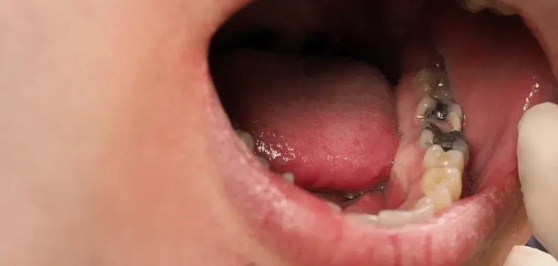
A decayed tooth may not initially look or feel unusual. Not every dark mark is decay either, and hidden decay may exist between teeth or beneath a restoration even when the tooth looks normal.
Possible signs include:
- A chalky white area
- Brown or black discolouration
- A rough area or visible hole
- Food repeatedly becoming trapped
- Sensitivity to cold, heat or sweetness
- Pain when biting
- Persistent toothache
- Bad breath or an unpleasant taste
- Swelling near the tooth
How Is Dental Decay Diagnosed?
A dentist assesses symptoms, risk factors and changes in the colour, texture or structure of the teeth. Diagnosis may involve visual examination, checking existing restorations and taking dental X-rays when appropriate.
Bitewing radiographs show the crowns of the upper and lower back teeth and may reveal decay between teeth or around restorations before it becomes visible.
The need for X-rays should depend on the person’s symptoms, age, dental history and individual risk.
Can Dental Decay Be Stopped or Reversed?
Early dental decay may sometimes be arrested or reversed while the enamel surface remains intact. Minerals from saliva, supported by fluoride, can return to weakened enamel through remineralisation.
The critical point is whether cavitation has occurred. Cavitation means that the tooth surface has physically broken down and formed a hole.
A non-cavitated lesion may respond to:
- Improved plaque removal
- Fluoride toothpaste
- Professional fluoride treatment
- Reduced sugar frequency
- Dry-mouth management
- Professional monitoring
Once part of the tooth has been physically lost, brushing and fluoride cannot rebuild its original structure.
Is Fluoride Good for Dental Decay?
Fluoride cannot fill an established cavity. Its greatest value lies in prevention and controlling early decay before the tooth surface breaks down. Fluoride has a well-established role in preventing dental caries and managing its earliest stages.
It can:
- Reduce mineral loss during acid attacks
- Support remineralisation
- Make enamel more resistant to acid
- Reduce bacterial acid production
Is Fluoride Safe?
Fluoride products should be used in age-appropriate amounts and according to instructions. Toothpaste should not be deliberately swallowed.
Excessive fluoride intake while permanent teeth are developing can cause dental fluorosis, a change in enamel appearance. Young children should therefore be supervised while brushing.
Adults cannot develop dental fluorosis because their permanent enamel has already formed. Prescription-strength products should still be used only as directed.
How to Stop Tooth Decay from Developing or Getting Worse
The best preventive approach combines plaque control, fluoride, sensible eating patterns and dental care based on individual risk.
Brush Twice Daily with Fluoride Toothpaste
Brush for approximately two minutes and clean every accessible surface. Use gentle movements rather than aggressive scrubbing, which can contribute to gum recession and tooth wear.
After brushing, spit out the excess toothpaste rather than immediately rinsing with water. If fluoride mouthwash is recommended, use it at a different time so it does not wash away the concentrated fluoride left by toothpaste.
Floss Your Teeth Every Day (Interdental Cleaning)
Toothbrush bristles cannot completely clean between neighbouring teeth. Plaque remaining there may contribute to interproximal caries, meaning decay between two teeth.
Guide floss gently between the teeth, curve it around each tooth in a C-shape and move it carefully along the side of the tooth and just below the gumline. Do not snap it into the gums.
Interdental brushes may be better for wider spaces, exposed roots, braces or bridges. Water flossers can help people who find conventional floss difficult. A dental professional can recommend the most suitable method.
Flossing cannot remove an existing cavity, but it helps disrupt plaque in areas that a toothbrush may miss.
Reduce Sugar Frequency
Helpful habits include:
- Keeping sweet foods and drinks to mealtimes
- Avoiding continuous sipping of sweetened beverages
- Choosing water between meals
- Checking products for hidden sugars
- Avoiding sugary food before bed
- Addressing persistent dry mouth
Ask About Dental Sealants
A dental sealant is a thin protective coating placed over the grooves of back teeth. It creates a smoother surface that helps prevent plaque and food from collecting.
Sealants are often used for children and teenagers, although some adults may benefit. They do not replace brushing, fluoride or interdental cleaning.
How to Get Rid of Cavities
An established cavity cannot be brushed away or permanently removed at home. Treatment depends on its depth and how much healthy tooth remains.
Condition | Possible treatment | Purpose |
Early lesion without cavitation | Fluoride and preventive management | Support remineralisation and stop progression |
Small or moderate cavity | Filling | Remove damaged tissue and restore the tooth |
Extensive structural damage | Inlay, onlay or crown | Rebuild and protect the remaining tooth |
Inflamed or infected pulp | Root canal treatment and restoration | Remove diseased pulp and preserve the tooth |
Tooth that cannot be restored | Extraction | Remove a severely damaged or infected tooth |
Not every dark spot requires drilling, and not every decayed tooth needs a crown. Treatment should be based on a clinical examination and diagnostic imaging when necessary.
Are There Natural Ways to Cure Tooth Decay?
Healthy habits can support saliva, limit acid attacks and encourage remineralisation of early enamel changes. However, no proven natural remedy can refill an established cavity.
Oil pulling, clove oil and salt-water rinses cannot rebuild lost enamel or dentine. Calcium-rich foods support general nutrition but cannot fill a hole. Reducing sugar can help prevent further acid attacks, but it cannot restore missing tooth structure.
“Natural” does not automatically mean safe or effective. Home remedies should never delay professional assessment of pain, swelling or suspected decay.
What Happens If Dental Decay Is Left Untreated?
Decay reaching the pulp may produce pulpitis, meaning inflammation of the nerves and blood vessels inside the tooth. Further progression can cause:
- Pulp necrosis, or death of the pulp
- Infection around the root
- Dental abscess
- Severe pain or facial swelling
- Tooth fracture
- Difficulty eating
- Tooth loss
- Movement of neighbouring teeth
- Disrupted sleep and daily activities
Seek prompt care for persistent toothache, pain when biting, swelling, fever or discharge. Swelling affecting the eye or neck, or difficulty breathing, speaking or swallowing, requires emergency medical attention.